Through the Looking Glass: Anesthesia and 3D Technology
SUMMARY: Peering into the inner workings of the heart is nothing new for poets and philosophers, but anesthesiologists have a seen a thing or two, as well. However, the standard TEE technology can only convey so much. A new TEE code allows you to bill for a much better view.
One of the more recent topics discussed and debated among theoretical physicists involves the nature and number of dimensions. In classical terms, there are three spatial dimensions: up and down, forward and back, and side to side; or, to put it briefly: height, depth and width. Time is considered the fourth dimension. With the publication of Albert Einstein's general theory of relativity, the world was introduced to the concept of "space-time" as a combined phenomenon. When the fabric of space-time is curved due to the intervening presence of celestial bodies, gravity results.
In the 1960s, a music group decided to name itself "the Fifth Dimension"—a reference to a more ethereal and unseen plane of existence beyond space and time. More recently, some physicists have postulated that there are as many as 11 dimensions. Rather than explore the legitimacy of, and evidence for, these imaginative ideas, our purpose herein is to focus more fully on the classically understood three dimensions of physical reality; or, to put it in its shorthand parlance: 3D. Specifically, we will be taking a look at how anesthesiologists can utilize and bill for 3D technology.
Beyond 2D TEE
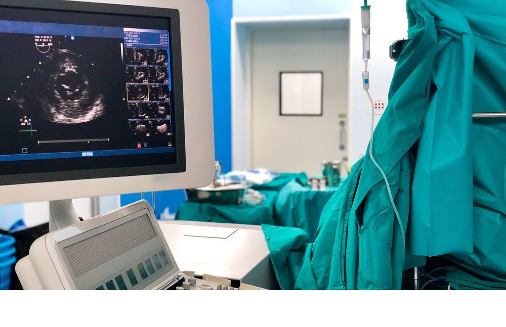
One of the innovative instruments created by the medical device industry for the cardiology community is the transesophageal echocardiography (TEE) probe and associated TEE imaging. This tool and technology are not only used by cardiologists but anesthesiologists, as well. While not every anesthesiologist is sufficiently credentialed or accredited in TEE, those who are, are well aware of how useful it can be in diagnosing or monitoring critically ill patients. These anesthesiologists are also aware that TEE typically comes in a 2D application. That is, the image reveals only height and width but no depth. The 2022 CPT coding manual actually describes TEE codes 93312 through 93318—the primary TEE codes—as follows:
An endoscopic ultrasound transducer is passed through the mouth into the esophagus and 2-dimensional images are obtained from the posterior aspect of the heart.
So, clearly, the typical TEE is only 2D. As great as TEE is, it's missing that third dimension, and this can limit the observer's perception. It reminds me of the example of Flatland—a mythical world where people only see and exist in two dimensions. Then a person from our 3D world accidently throws a ball across the Flatland sky. The Flatlanders can only see a circle. They are unable to perceive the sphere. It's a bit of a handicap, like a 2D TEE. However, there are 3D options when it comes to TEE services, including a new code for 2022.
Code 93319 is a new entry in the TEE code set found within the CPT manual for this year. It contains the following descriptor:
3D echocardiographic imaging and postprocessing during transesophageal echocardiography, or during transthoracic echocardiography for congenital cardiac anomalies, for the assessment of cardiac structure(s) (eg, cardiac chambers and valves, left atrial appendage, interatrial septum, interventricular septum) and function, when performed (List separately in addition to code for echocardiographic imaging)
That last parenthetical phrase of the code descriptor indicates that this is an add-on code, meaning that it would be listed in addition to the applicable 2D code performed on the case (e.g., 93312, 93314, 93315 and 93317).
Implications and Implementation
In practical terms, what this add-on service means is that, during the same session as the primary echocardiography service, the provider uses a 3D–capable machine to perform 3D imaging. The provider can see the images in real–time and can also use postprocessing software for further studies, such as reconstruction and measurement.
When the patient is appropriately prepped and possibly anesthetized, the provider performs 3D echocardiographic imaging at the same session as the primary service for transesophageal echocardiography (with the probe in the esophagus), transesophageal echocardiography for congenital cardiac anomalies, or transthoracic echocardiography (with the probe on the chest area) for congenital cardiac anomalies. The provider uses the probe (transducer) of a 3D–capable machine to send sound waves that bounce off body tissues to make echoes (ultrasound). The probe receives the echoes and sends them to a computer, which uses the signals to create an image.
The provider can view the 3D imaging in real time to assess one or more cardiac structures, such as the cardiac chambers and valves, left atrial appendage, interatrial septum, and interventricular septum. The provider also may assess function. Postprocessing software allows the provider to reconstruct images, take measurements and perform other functions after acquiring the ultrasound images and data.
Reimbursement Ramifications
This service was previously coded with CPT code 76376, but the new code was established to capture the additional physician work and practice expenses associated with real-time 3D echocardiography.
From time to time, we have made reference in these alerts to the National Correct Coding Initiative (CCI), a Medicare program that determines bundling rules, i.e., what services are subsumed into a comprehensive coded service. There is a column 2 CCI edit rule for new code 93319 and existing code 93325 (color flow velocity mapping). This indicates you may not report these two codes together on a claim. Previously, when 93325 was reported with 76376 (which 93319 now replaces), there was no CCI edit between the two. That is, you could bill the two services together. That is now no longer the case. The 2022 CPT manual also contains a stipulation that states, "Do not report 93319 in conjunction with 76376, 76377, 93325,93355."
The total facility relative value unit (RVU) value of CPT 93319 is 0.73, and the total non-facility RVU value is 1.79. The previous code, 76376, has a total facility RVU of 0.28, and 93325 (which previously could be coded together with 76376) has a total facility RVU of 0.09. So, the single code, 93319, is valued higher than the two previous codes that were allowed together. Therefore, from a reimbursement perspective, the new coding option availed by 93319 is an overall positive for those anesthesiologists utilizing 3D imaging.
It isn't every day one gets to see the inner workings of the heart with such clarity and precision. When one does, it's good to know that he or she is appropriately compensated. If you have questions about TEE code 93319, please contact your account executive or reach out to us at info@anesthesiallc.com.
With best wishes,
Tony Mira
President and CEO